 |
|
The ultimate resolution of a Fresnel zone plate is determined by the width of the outermost zone. The focal spot is surrounded by rings of intensity (secondary maxima) that blur the images obtained in X-ray microscopy and scanning spectroscopy. These limitations can be overcome by using a large number of
appropriately distributed pinholes instead of rings as the diffracting elements.
To obtain a distinct first-order focus, the pinholes have to be positioned such
that the optical path length from the source via the center of the pinholes to
the focal point is an integral number of wavelengths. |
|
|
|
Fig. 1 Photon sieve. Tens of thousends of pinholes are
arranged to diffract light or X-rays to a small focus.
|
|
Furthermore, with photon sieves, the unwanted ring-like
secondary maxima generated by zone plates can be
suppressed. For a zone plate each ring contributes equally to the amplitude in
the focus. This contribution drops abruptly to zero beyond the outermost ring
which leads to strong intensity oscillations in the diffraction pattern. With a
photon sieve the number of pinholes per ring can be readily adjusted to yield a
smooth transition which minimizes the secondary maxima. |
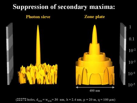 Fig. 2 Intensity distribution on the focal plane of a photon sieve compared to a zone plate. Smallest structure size for both optical elements is 30 nm. For the photon sieve, the suppression of the secondary maxima is evident. This provides sharper images. |
|
The development of fourth-generation synchrotron light sources that deliver coherent radiation in the soft X-ray regime in combination with photon sieves will provide a wealth of new opportunities in X-ray microscopy, spectroscopy and lithography. Photon sieves for use at the Hamburg Free-Electron Laser facility (HASYLAB) are currently being developed. |
 Fig. 3 Schematic of a nanospectroscope capable of recording high-resolution images revealing chemical composition, morphology, and the electronic properties of materials in the nanometer regime. |